

Cellular Biology
Autophagy and Cellular Recycling: What Researchers Measure
Autophagy delivers cytoplasmic material to lysosomes for degradation and recycling. Explore the experimental methods, interpretation limits and open questions that shape this field.
7 November 2026 · 5 min read · Novaryn Labs
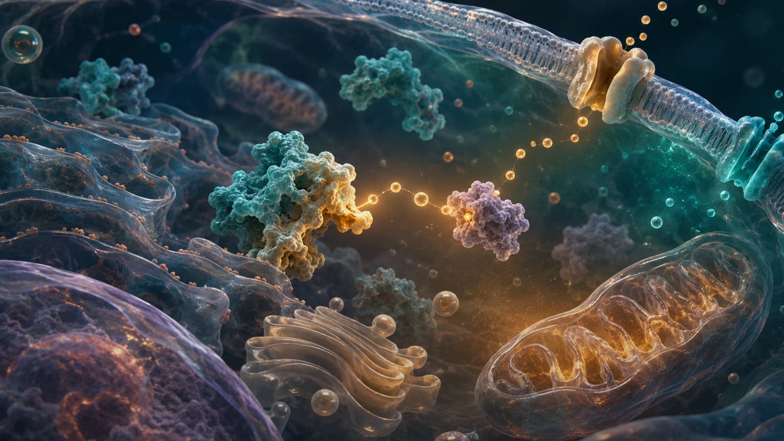
Autophagy delivers cytoplasmic material to lysosomes for degradation and recycling. Macroautophagy forms double-membrane autophagosomes, while other routes use different delivery mechanisms.
The important research question is not whether autophagy and cellular recycling can be attached to a broad biological claim. It is which molecular event was measured, in what model, over what timescale and with which controls. This guide develops that distinction so readers can move from an attractive mechanism to a testable interpretation.
Novaryn Labs presents this material as scientific education for laboratory and analytical work. It does not provide personal-use instructions, medical guidance or a substitute for reviewing the complete methods and limitations of the cited literature.
The core scientific idea
Autophagosome formation begins with regulated membrane assembly and cargo selection before fusion with the lysosomal compartment.
LC3-related measurements can report autophagosome abundance, but abundance rises when formation increases or when downstream clearance is blocked.
Autophagic flux therefore compares pathway behaviour with and without a lysosomal block rather than relying on one static marker.
Together, these points show why autophagy and cellular recycling cannot be reduced to one marker or diagram. A convincing experiment defines the molecular species, the biological or analytical context and the endpoint before interpreting a change. That discipline also makes apparently conflicting findings easier to compare.
How researchers investigate autophagy and cellular recycling
No single method captures identity, mechanism and function at once. Strong projects combine measurements that fail in different ways, then ask whether the results support the same explanation.
LC3 and p62 measurements paired with flux controls
LC3 and p62 measurements paired with flux controls can establish a defined part of the autophagy and cellular recycling question. Researchers should state the sample, comparator, timing and acceptance rule before collecting data. Replication across independent preparations helps distinguish a repeatable signal from instrument drift or one unusual sample.
Microscopy of autophagosome and lysosome dynamics
Microscopy of autophagosome and lysosome dynamics adds a second view of the same system. Its strongest use is alongside an orthogonal measurement that relies on a different physical or biological principle. Agreement is informative; disagreement is equally useful because it reveals where identity, timing or model assumptions need closer examination.
Genetic perturbation of core autophagy machinery
Genetic perturbation of core autophagy machinery connects the molecular observation with a broader experimental outcome. Controls should include the untreated system, an appropriate positive or process control and checks for assay interference. The method should be interpreted within its validated range rather than extended beyond the data.
What the evidence can and cannot establish
- Interpretation limit one: marker interpretation varies by tissue and assay
- Interpretation limit two: more autophagosomes do not necessarily mean more recycling
- Interpretation limit three: acute stress responses may differ from long-term adaptation
For autophagy and cellular recycling, these limits do not make the research uninformative. They define the boundary of the conclusion. Evidence about a purified molecule can be strong evidence about that molecule, while remaining incomplete evidence about a cell. A cellular response can be reproducible without predicting a whole organism. Human observations can identify an association without resolving the molecular cause.
The most reliable reading of autophagy and cellular recycling therefore follows an evidence ladder. First confirm identity and measurement performance. Then ask whether perturbing the proposed mechanism changes the endpoint. Finally, look for replication in models that add relevant complexity. Each step should preserve the uncertainty of the step before it.
Current research questions
Is a marker change caused by formation or impaired clearance?
Answering this requires a design that separates the proposed mechanism from correlated changes. Time-resolved measurements, matched controls and a clear definition of the primary endpoint are more informative than collecting many endpoints without a decision rule.
Which cargo is being selected under the tested condition?
This question often needs more than one model. A reductionist assay can isolate mechanism, while a more complex system can test whether that mechanism remains important when feedback, metabolism and competing pathways are present.
Frequently asked questions
What is autophagy and cellular recycling?
Autophagy delivers cytoplasmic material to lysosomes for degradation and recycling. The term is useful only when the molecular process and experimental context are stated clearly.
Why do researchers study it?
Researchers study autophagy and cellular recycling because it connects measurable molecular events with wider questions in cellular biology. It also provides a way to test how structure, timing or analytical conditions alter an observed result.
How is it measured?
Common approaches include LC3 and p62 measurements paired with flux controls, microscopy of autophagosome and lysosome dynamics and genetic perturbation of core autophagy machinery. These methods answer different questions and are strongest when used together.
What is a common interpretation mistake?
A common mistake is assuming that marker interpretation varies by tissue and assay. A study can be technically sound while supporting a narrower conclusion than its headline suggests.
Does laboratory evidence prove a human outcome?
No. Molecular, cell and animal studies can establish mechanism within their models, but translation requires appropriately designed human research. Even human studies must be interpreted within their participants, endpoints and follow-up period.
What should be checked before comparing two studies?
Check the molecular material, model, comparator, timing, primary endpoint and analytical method. Differences in any of these can explain why two studies that appear to address autophagy and cellular recycling reach different results.
Related research
Continue with Understanding Mitochondrial Function: Energy, Signals and Measurement and The Biology of Cellular Ageing: Mechanisms, Markers and Limits. These guides approach the same research area from complementary structural, biological or analytical perspectives.
Scientific references
- Liu S, Yao S, Yang H et al. (2023). Autophagy: Regulator of cell death. Cell death & disease. DOI: 10.1038/s41419-023-06154-8.
- Mizushima N, Komatsu M (2011). Autophagy: renovation of cells and tissues. Cell. DOI: 10.1016/j.cell.2011.10.026.
- Nixon RA, Rubinsztein DC (2024). Mechanisms of autophagy-lysosome dysfunction in neurodegenerative diseases. Nature reviews. Molecular cell biology. DOI: 10.1038/s41580-024-00757-5.
Questions about anything in this guide?
Message the team on WhatsApp for product, batch or documentation questions. We can't give dosing or medical guidance.
For laboratory research use only. Not for human or veterinary consumption. Nothing in this article is medical advice.

